- Publikationen ca: 1
- Fragen & Antworten
Jasper Frohn
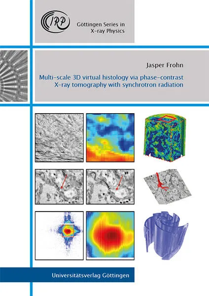
Multi-scale 3D virtual histology via phase-contrast X-ray tomography with synchrotron radiation
To this day, the standard method for investigating biological tissue with cellular resolution is the examination under a light microscope, first denoted as histology by Karl Meyer in 1819. Despite the enormous success and importance of histology, it has two major disadvantages.
